Biophoton research in blood reveals its holistic properties
V.L. Voeikov*#, R. Asfaramov,* E.V. Bouravleva*, C.N. Novikov*, N.D. Vilenskaya*.
#International Institute of Biophysics, Neuss, FRG, *Faculty of Biology, Moscow State University
Abstract
Monitoring of spontaneous and luminophore amplified photon emission (PE) from non-diluted human blood under resting conditions and artificially induced immune reaction revealed that blood is a continuous source of biophotons indicating that it persists in electronically excited state. This state is pumped through generation of electron excitation produced in reactive oxygen species (ROS) reactions. Excited state of blood and of neutrophil suspensions (primary sources of ROS in blood) is an oscillatory one suggesting of interaction between individual sources of electron excitation. Excited state of blood is extremely sensitive to the tiniest fluctuations of external photonic fields but resistant to temperature variations as reflected in hysteresis of PE in response to temperature variations. These data suggest that blood is a highly cooperative non-equilibrium and non-linear system, whose components unceasingly interact in time and space. At least in part this property is provided by the ability of blood to store energy of electron excitation that is produced in course of its own normal metabolism. From a practical point of view analysis of these qualities of blood may be a basement of new approach to diagnostic procedures.
Introduction
Ultra-weak photon emission (PE) in the optical spectral range from many cells is commonly thought to represent random imperfections accompanying normal physiological processes of oxygen consumption, and no biological function is usually ascribed to it. However evidence is accumulating that electron excited species are regular products of biochemical processes and that energy of their relaxation may be used by living systems in different ways, such as provision of excitation energy for endergonic chemical reactions, photomodulation of enzyme activities, ets. . Energy of electron excitation may be transmitted from the sites of its generation (donors) to the sites of its utilization (acceptors) both by radiation-less as well as by radiative mechanisms .
The major sources of electron excitation in living systems are the reactions with the participation of reactive oxygen species (ROS), in particular, the reactions of superoxide anion radical (O2• ⎯) recombination, yielding hydrogen peroxide (H2O2) and singlet (electronically excited) oxygen (*O2), reactions of H2O2 reduction to water and oxygen or of oxidation by it of chlorine ions, reactions of direct oxidation of carbonyl compounds with oxygen from which reaction products in a triplet excited state arise, etc. These reactions may go on non-enzymatically or be enzyme catalyzed and are under strict external regulation besides a probable ability to be self-regulatory . A substantial portion of oxygen consumed by aerobic organisms is permanently used for generation ROS, thus, electronic excitation should also be permanently generated.
Among the mostly intensively studied biological sources of PE are stimulated neutrophils and other phagocyting cells. They react to multiple stimuli by a respiratory burst (RB) – strong intensification of ROS production followed with PE . As intensity of this emission is very low, PE indicators, such as luminol or lucigenin are introduced into a cellular suspension to increase quantum yield . Luminol and lucigenin are known to be indicators of different oxygenation activities. Lucigenin is regarded as a relatively selective probe for O2• ⎯, while luminol is less specific and reports of a variety of reactive oxygen species (H2O2, ClO⎯, OH•, etc.) production . Non-diluted blood is a priori considered to be completely non-transparent for visible light because of a very high hemoglobin` content, and it is practically never used for PE studies. However, here we show that significant photon emission can be registered from non-diluted human blood, both in a resting state in the presence of lucigenin and especially when agents inducing RB of neutrophils are added to it. Patterns of PE revealed due to continuous monitoring of it allowed to expose peculiar systemic properties of blood.
Materials and methods
Reagents
All reagents unless otherwise specified were obtained from Sigma Chemical Co., USA. Stock solution (10-1 M) of luminol was prepared in analytical grade dimethyl sulfoxide. It was diluted 50-fold in saline just before use and added to a blood sample to a final concentration of 10-4 M. Stock solution (10-2 M) of lucigenin was prepared in saline (0.9% sodium chloride solution). It was added to a blood sample to a final concentration of 10-4 M. Zymosan was opsonized with human blood serum by a routine procedure and was added to blood to a final concentration of 0,1 mg/ml.
Preparation and treatment of blood samples
Blood from healthy volunteers was obtained by venous puncture between 9 and 11 hours a.m. and was stabilized by heparin or sodium citrate. Blood was kept in 5 or 10 ml plastic disposable syringes without air bubbles at 20 oC or at 4 oC if it was stored for more than 6 hours. In these cases blood was kept at room temperature for 1 hour before the measurements. For experiments described in this paper blood of healthy donors (males, 20 – 51 years old) was used. Individual differences in PE kinetic curves progression, PE maximal intensity were noted, though they were reproducible in experiments with blood of each particular donor. General trends of PE from blood exemplified at figures presented in the paper were typical for blood of all the donors.
The ability of neutrophils to reduce nitro-blue tetrasolium (NBT), expressed as percent reducing neutrophils was evaluated by a common method . In brief an aliquot of blood (50 mkl) was taken from a blood sample, mixed with 50 mkl of 0,1% NBT solution in 0,15 M Na-P buffer (pH 7,2), the mixture was incubated at 37 oC for 20 min and at 20 oC for 20 min. Blood was smeared on a slide, dried out, fixed with methanol for 10 min. Slides were dyed with methylene green. 100 neutrophils were counted at each slide. NBT test was considered positive when dark blue diformasan granules (the product of NBT reduction) occupied from 5 to 100% of neutrophils cytoplasm. All the cells were grouped in 6 ranks according to their activity: range 1 — 0%, range 2 — 5-7%, range 3– up to 30%, range 4 — 30-50%, range 5 — 50-90%, range 6 — 100% of cytoplasm is filled with reduced diformasan granules.
Detection of photon emission
PE from blood was registered either in a liquid scintillation counter Mark-II (Nuclear-Chicago, USA), equipped with photomultipliers EMI 9750QB/1 or on a single photon counter equipped with PMT type EMI 9558 QA, cooled to –200С +- 0,2 0С . Mark II counter was used in the mode of single photon counting (out-of-coincidences mode) in a tritium window. The measurements were performed at room temperature (19-21 oC). PE was recorded as counts per 0.2 or 0.1 min. 1 ml Eppendorf polyethylene test tubes were used as blood containers. Test tubes were fixed in empty standard borosylicate glass vials for liquid scintillation counter in one and the same position. Vials and test tubes having short decay time of own luminescence after insertion into the counting chamber were selected. Dark counts with an empty test-tube in a counting chamber varied in the range of 40-50 counts/sec. All the operations were performed at dim ambient illumination. Sequence of addition of blood, luminol, lucigenin and zymosan are described in figure legends. Other experimental details are described in the section “Results”. Single photon counter7 was used in experiments where temperature dependence of PE from blood was measured. Blood was placed in standard disposable transparent plastic cuvettes for spectrophotometers, and a cuvette was fixed in a copper container with one transparent wall and equipped with the Peltier element for its heating and cooling. A thermistor coupled to the Peltier element was inserted to blood and using a special software temperature in blood could be changed and PE and blood temperature were simultaneously recorded.
Results
Photon emission from blood and its dependence on oxygen.
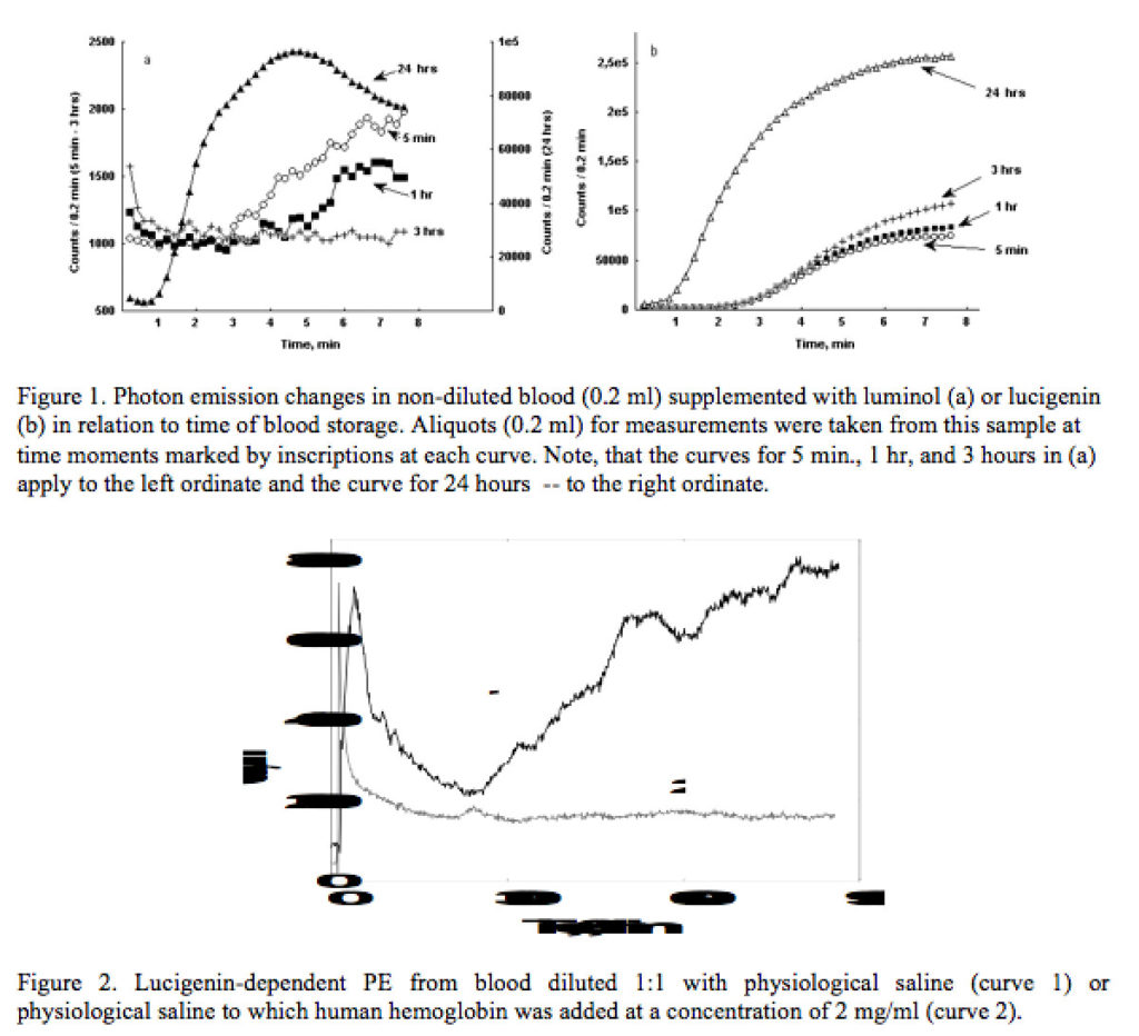
Addition of either lucigenin or luminol to blood as soon as 5 min after it had been taken out by a finger puncture or elbow vein drainage is followed with an increase of PE in the absence of RB stimulants (Figure 1). Fresh blood response to lucigenin was much more pronounced than that to luminol. During the first hours of blood storage after its withdrawal luminol-dependent PE (LM-PE) level was decreasing, but addition of luminol to 1 day old blood resulted in rapid and strong elevation of LM-PE (Fig 2a). Unlike complex pattern of LM-PE changes during blood storage, lucigenin-dependent PE (LC-PE) was not decreasing, but was rather gradually increasing during blood storage. Similar to LM-PE strong enhancement of LC-PE was observed in 1-day old blood (Fig 2b). Blood even in a resting state (without addition of inducers of RB) to which lucigenin was added continued to emit photons for many hours indicating that the process of ROS production and generation of electron excited species persistently proceeds in it.
The very fact that pronounced PE may be registered from non-diluted blood — a highly opaque liquid due to very high concentration of hemoglobin — indicates that hemoglobin packed in erythrocytes does not quench efficiently PE. However, if free hemoglobin is added to blood at a concentration of only 0,5% of already present in it, LC-PE practically disappears (Figure 2). Taking into account that concentration of hemoglobin in erythrocytes may reach as high value as 35-40% (hemoglobin can not reach such high concentration in a free solution) one may suggest that hemoglobin in erythrocytes is present in a liquid crystalline state. In such a form it may provide transfer of excitation energy over long distances without its dissipation, unlike hemoglobin in a solution that absorbs and dissipates energy of electron excitation.